


When we talk about exophthalmos, we refer to an excessive protrusion of the eyeball on one or both sides.
The most frequent cause of exophthalmos is thyroid orbitopathy, a particular condition of transformation of the eyes that occurs as a consequence of an autoimmune disease that affects the thyroid and manifests with excessively protruding eyes, eyelid swelling, and excessive eyelid opening or eyelid retraction.
Excessive eyelid opening, eyelid retraction, is often associated with exophthalmos, which contributes to increasing the effect of the eyeball protrusion.
In some patients, an improvement of the ocular condition can occur with appropriate therapy for the thyroid, but often the damage is permanent and requires a surgical solution.
Treatment of thyroid orbitopathy
This disease typically presents two phases:
- The active, inflammatory phase: it is the first phase and has a variable duration from 6 to 18 months on average and tends to resolve spontaneously. In this phase the patient must be followed by the endocrinologist to achieve a stabilization of the thyroid function, which can be reached with medical therapy (Tapazole), suppressive (radioiodine) or surgical (thyroidectomy).
At the same time, ocular disorders, which must be evaluated and managed together with the ophthalmologist, primarily include the use of cortisone, which can be administered intravenously, orally, or with local infiltrations made in the lower eyelid, which allows to reduce systemic complications and maximize the ocular therapeutic effect. - The quiescent, fibrotic phase: it is the phase that follows the end of the inflammation and is characterized by a complete stabilization of ocular signs and symptoms, without signs of inflammation. The eye is white, the swelling and redness are gone, but the damage occurred in the active phase, which did not improve spontaneously or with cortisone, is permanent.
In this phase, medical treatment is ineffective and even if thyroid function is normalized, no further improvement can be expected. It is in this phase that the surgeon comes into play, who, having verified that the condition has been stable for at least 6–8 months, can offer the patient a complete and effective reconstructive aesthetic rehabilitation.
Surgical therapy of exophthalmos
Orbital decompression is the procedure performed to correct the excessive protrusion of the eyeballs (exophthalmos) using minimally invasive techniques, without visible scars and in a day surgery setting.
The technique involves the enlargement of the bony orbital space thanks to the removal of the two thinnest orbital walls that border the nasal sinuses.
A second surgery may be necessary to correct upper and lower eyelid retraction, possibly combined with the removal of excess fat, if needed.
At the end of the surgical phase, the patient should appear as close as possible to the pre-disease state.

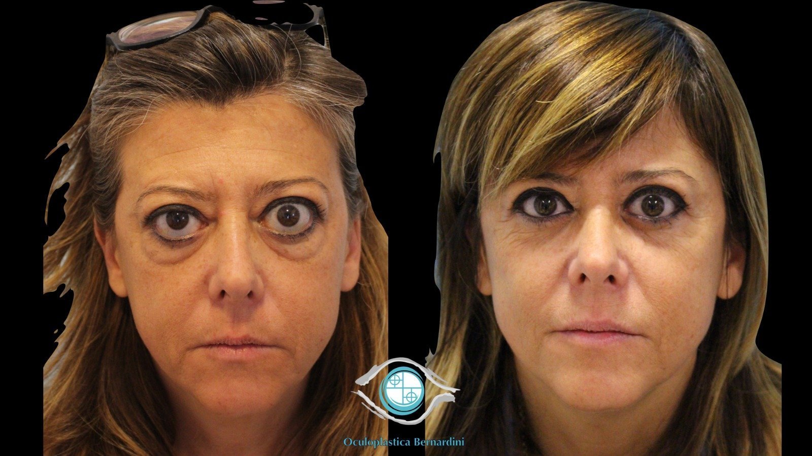
Photos before and after correction of exophthalmos

Right: post-operative appearance 2 months after orbital decompression and associated blepharoplasty..


Here you can find more before and after photos of cases treated by Prof. Francesco Bernardini

The recommendations of the American Society of Ophthalmic Plastic and Reconstructive Surgery
There are many causes of ptosis including age-related weakening of the muscle, congenital weakness, trauma, or sometimes neurologic disease. As we age, the tendon that attaches the levator muscle, the major muscle that lifts the eyelid, can stretch and cause the eyelid to fall.
This represents the most common cause of a droopy eyelid. Ptosis may also occur following routine LASIK or cataract surgery due to stretching of the muscle or tendon. Children may be born with ptosis or may acquire it due to trauma or neurologic reasons.
