Injectable Techniques with HA (Hyaluronic Acid) for Non-Invasive Eye Rejuvenation
Scientific studies have shown that the periocular area is central to the aesthetic appeal of the face. The first signs of aging are perceived around the eyes, and 80% of patients between the ages of 45 and 50 seek eye rejuvenation or “beautification” as their initial treatment.
How the Eye Area Ages
Facial volume loss, caused by the atrophy of various fat compartments, leads to the appearance or worsening of imperfections, especially noticeable at the lower eyelid and the cheek below it. This area, known as the “eyelid-cheek junction” in English literature, is a delicate transition zone between the lower eyelid and the cheek. With aging, the lower eyelid lengthens, eye bags become prominent, the orbito-malar groove deepens, and the cheeks appear to sag.
Additionally, skin laxity and the development of static and dynamic wrinkles occur. The lower eyelid rounds out, and the lateral canthus moves medially. These changes are vividly depicted in the portraits of Rembrandt, specifically in the portraits of an “Old Man” and a “Young Woman.”

Aging of the Eye Area
Constitutional Dark Circles: In addition to the static and dynamic signs of aging, there are congenital imperfections that can impact the appearance of the eyes and may have always been present. Patients often describe these as “eye bags,” “dark circles,” or “puffy eyes,” often in varying combinations.
The complexity of these issues lies in the intricate anatomy of the eyelid, which is separated from the cheek by strong orbital ligaments, forming the so-called “tear trough” between the eye and the nose, and the “orbito-palpebral sulcus,” also known as dark circles.
The goal of filler treatments is to correct the transition between the eyelid and the cheek, addressing all imperfections in the infraorbital region rather than focusing solely on the tear trough.
The injector must always be mindful of the local anatomy, treating the injection site as if the needle or cannula tip were an ultrasound probe guiding them appropriately. Especially in the treatment of the infraorbital area, an in-depth understanding of anatomy is essential for achieving aesthetically pleasing results and avoiding complications.
A treatment that focuses solely on filling the tear trough, whether with a needle or cannula, is often inadequate and unsatisfactory. This approach fails to address the broader infraorbital region and neglects the root of the problem.
Advanced Techniques
Drawing from surgical principles, we have developed an injection technique specifically for the infraorbital region, taking into account the physical properties of different fillers.
Broadening our perspective and moving away from a “fill-the-hole” approach, we can expand the scope of filler treatments. This allows us to address not only the main concern (tear trough deformity) but also achieve a “surgical-like” result through a non-invasive technique. This approach corrects volume deficits, minimizes eye bags, supports the eyelid, and improves skin quality.
By combining different types of fillers—such as a “heavy” filler for lateral lifting and central support and a “light” filler designed for the delicate eye area—it is possible to refine transitions and achieve a “Photoshop effect.”

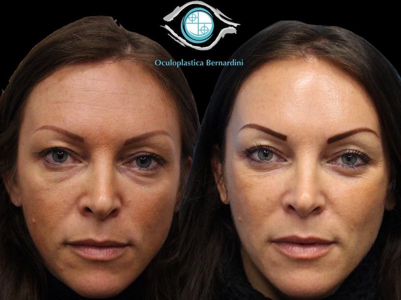
the photo demonstrates the right side post-treatment and the untreated left side, highlighting the immediate effects on eye bags, dark circles, and skin quality
The benefits of the technique I use include reduced risk of complications and broader treatment indications, with highly satisfying outcomes in most cases, both subjectively and objectively.
In my practice, medical treatments have expanded the possibilities for addressing dark circles, tear troughs, eye bags, and skin quality, reducing the need for surgical interventions. However, surgical treatments remain necessary for addressing excessive medial eye bags, festoons, and significant eyelid laxity or malpositions.
Anatomical Considerations
As shown in the image, the following aesthetic defects can be identified in both younger and older individuals:
- Tear Trough Deformity (Yellow)
- Orbito-Malar Groove (Green)
- Eye Bags

The specificity of these issues lies in the complex anatomy of the eyelid, which is separated from the cheek by strong orbicular ligaments. These form the so-called ‘tear trough’ between the eye and the nose, as well as the ‘orbito-palpebral sulcus,’ also known as dark circles.
The goal of filler treatment is to correct the transition between the eyelid and the cheek, addressing all imperfections in the infraorbital region rather than focusing solely on the ‘tear trough.’
The injector must always keep the local anatomy in mind, treating the tip of their needle or cannula as if it were an ultrasound probe guiding their technique appropriately.
Nowhere is anatomical knowledge more essential than in treating the infraorbital area, where it is crucial for achieving aesthetically satisfying results and avoiding unpleasant complications.

From oculoplastic surgery, we have learned that the ORL and TTL ligaments are firmly attached to the orbital rim bone, and lifting them requires a direct incision at their insertion point (Figure 7). This explains why it is challenging to stretch these ligaments simply by “filling” them with fillers, as shown in Figure 6.
For this reason, the technique of correcting volume defects by filling them with a needle or cannula—the most common method of treating the tear trough—is often considered inadequate and a source of dissatisfaction among patients, as well as potential issues. Moreover, as shown in Figure 3, focusing solely on the tear trough is a significant limitation, as it addresses only the medial third of the defect.
Another issue with directly treating the tear trough is that this approach requires substantial quantities of filler to “lift” the TTL. For reasons that remain poorly understood, this area is particularly prone to complications from fillers.
The risk of complications from fillers in the periocular region mainly arises from the development of chronic eyelid and pre-malar edema, which is a source of dissatisfaction for patients and difficult for injectors to manage. It is evident that minimizing the amount of material injected in this area is crucial to reducing the risks of both short-term and long-term complications.

G-Point Lift: Combining Medicine and Aesthetic Surgery

From surgery, we have learned that for the rejuvenation of the infraorbital region, suspending the orbicularis muscle during lower blepharoplasty results in an almost complete correction of the eyelid-cheek junction (Figure 7).
When this is combined with the transposition of palpebral fat to the central-medial area, the outcome is highly effective (Figures 8-9).
Aesthetic Surgery Journal 2015
Surgery and fillers share several anatomical concepts: the need for structural lifting, the existence of a maximal lift point (“G-Point”), central support indications (DMFC), and the opportunities to smooth transitions.
By applying surgical concepts to fillers, we have developed a specific injection technique for the infraorbital region, tailored to the physical characteristics of different fillers.
If we broaden our perspective and move away from the defect-focused “gap-filling” approach, we can expand the scope of filler treatments beyond just the primary issue—the tear trough. This enables us to achieve a “surgical-like” result with a non-invasive technique, addressing volume deficits, minimizing eye bags, supporting the eyelid, and improving skin quality.
By combining different types of fillers, such as a “heavy” filler for lateral lifting and central support, along with a “light” filler specifically designed for the eye area, it becomes possible to refine transitions and achieve a “Photoshop effect.”
The G-Point is determined by tracing a bisector from Hinderer’s lines (1: from the tragus to the nasal wing; 2: from the outer corner of the eye to the corner of the mouth). This bisector ascends towards the temporal fossa and intersects with a tangent originating from the outer corner of the eye.
This point, used in both surgery and medicine, is referred to as the “esthetic G-Point.” It is the first point of injection, where a deep macro-bolus of a high G-Prime filler is placed to achieve the lifting effect. Centrally, at the apex of the V-deformity, another deep macro-bolus is injected to provide central support.
Figure 10

Figure 11
Preliminary Study:
Over the past two years, I have treated the infraorbital area of 163 patients using the G-Point Lift technique. Specifically, for high G-Prime fillers, I alternated between Ultradeep or RHA4 (Teoxane) depending on various local characteristics such as skin thickness, and for low G-Prime fillers, I used Redensity II (Teoxane), particularly suited for the delicate lower eyelid area.
Results:
Objective analysis six months post-treatment demonstrated that 100% of patients showed significant improvement, rated between 3 and 4 on a 4-point scale.
Subjectively:
- 73% of patients rated themselves as “very improved” (4),
- 22.7% as “improved” (3),
- Only 2 patients (1.23%) reported no change (2),
- No patient rated themselves as worsened (0).
Complications:
Only 5 patients (3%) reported localized swelling, visible at rest or with facial expressions. These were successfully treated with hyaluronidase.
Conclusions:
This personal study has demonstrated that applying surgical concepts to aesthetic medicine allowed me to avoid direct treatment of the tear trough and instead treat the entire infraorbital region in a more comprehensive and harmonious way. This approach also reduced the amount of filler injected directly into the medial third.
The advantages of this technique include:
- Reduced risk of complications,
- Expanded treatment indications,
- Consistently satisfactory results in most cases, both subjectively and objectively.
In my practice, medical treatments have expanded the scope for addressing dark circles, tear troughs, and eye bags, while also improving skin quality. This has led to a reduction in strictly surgical indications, which remain necessary for treating excessive medial eye bags, festoons, significant eyelid laxity, and malpositions.